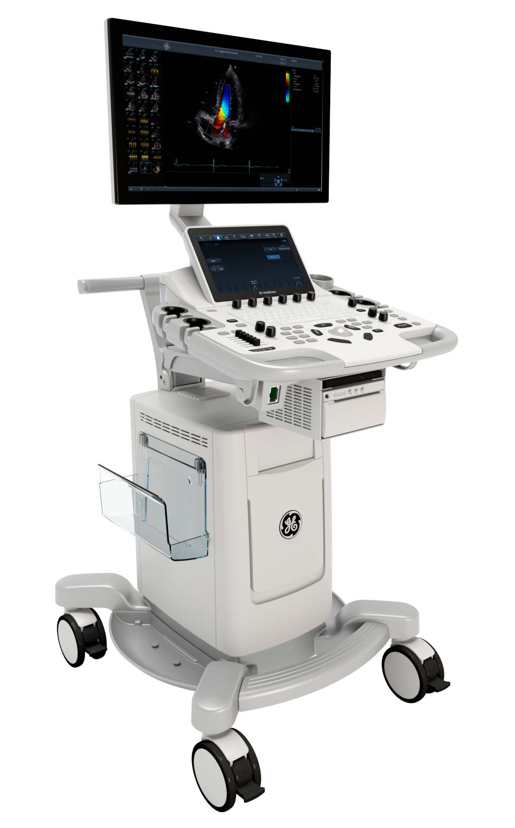
ViVid T8 心臟超音波

 |
Ergonomics & Workflow Modern. Efficient. Familiar.
|
Image quality and functionality
A feature-rich system with established Vivid software, uses a range of advanced quantitative tools to enhance its imaging in the cardiology field.
| Tissue Velocity Imaging* – Acquires dynamic myocardial data for quantifiable measurement of left ventricular function. | SmartStress* - Automatically adjust scan parameters to optimize workflows, improve reproducibility, and increase diagnostic confidence. |
 |
|
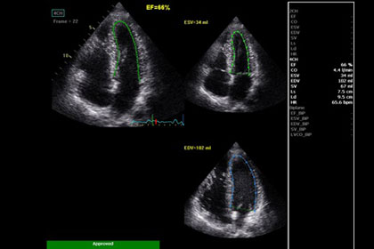
| AutoEF* – Automated assessment of left ventricular ejection fraction using an automated speckle tracking ROI tool. | Tissue Tracking*/Tissue Synchronization Imaging* – Provides additional image quality enhancement for evaluation of asynchronous cardiac wall motion. |
| Automated Function Imaging* – Evaluates and quantifies left ventricular wall motion at rest and calculates parameters to describe left ventricular myocardial function. Nearly 400 papers have been published on the AFI, whose proven reliability and reproducibility have also been confirmed by GE Healthcare. | Strain/Strain Rate Imaging* – Assists in visual and quantitative detection of myocardial segment dysfunction and may aid in the assessment of regional systolic function in ischemic heart disease. |
Sonography
The flexible Vivid T8, with its outstanding image quality in the multifunctional range, offers you options that allow you to customize the system to meet the diverse needs of your facility.
 |
|
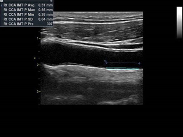
| Auto IMT* – Automatic borderline detection for intima-media thickness and automatic performance of all required measurements. | B-Flow* – Advanced spatial and temporal resolutions to support assessment of blood flow and vessel wall structure without the typical limitations of a Doppler technique. |
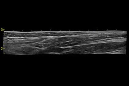
| Virtual Convex – Extends the field of view when using linear transducers. | Blood flow imaging* – Improved visualization of blood flow dynamics using a signal processing algorithm to visualize blood flow data. |
 |
|
| LOGIQview* – Increases the field of view for large organs that typically cannot be displayed in a single image. |