Voluson P8
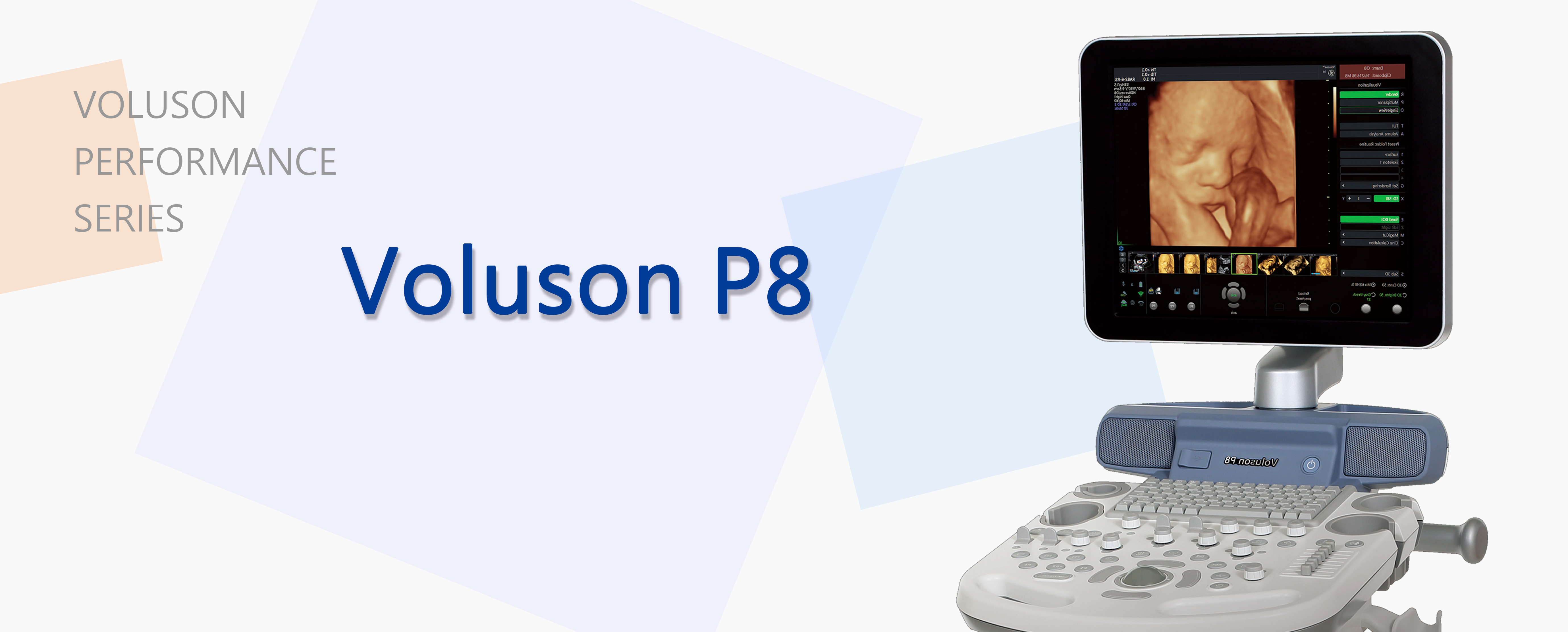
Voluson™ technology has established a high standard for 2D and 3D imaging — the Voluson Performance Series continues the tradition. With your VP8, you can count on exceptional images in all applications, from routine OB evaluations and gynecological studies to cardiac, abdominal, vascular, and small parts scanning.
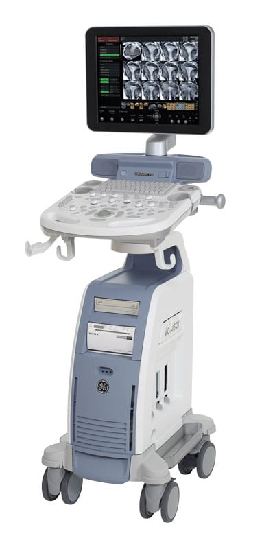
A sleek, ergonomic lightweight console designed for ease of use and comfort with integrated 17” flat-screen LCD monitor for stress free scanning.
|
|
Features included as standard:
|
Simply place the ultrasound probe to directly obtain the image information you need, without time-consuming modifications of settings. With the Voluson P8 system, you can acquire excellent clinical images across a broad range of patients, quickly and easily.
- Export PDF reports with images and graphs at the touch of a button.
- Advanced colour Doppler – For distinct imaging of adjoining vessels and flows.
- Exceptional penetration with consistent image quality – Delivers reliable results even with difficult-to-image patients.
- One-touch image optimisation – For fast and efficient imaging.
Easy 3D imaging
3D/4D imaging expands the spectrum of your diagnostic capabilities by providing additional anatomical perspective.
- Obtain planes not attainable in 2D imaging – for more accurate assessment.
- Analyse volume data during the examination or retrospectively after completion of the examination – for efficient patient management.
- Various capabilities for processing of volume data and displaying in form of single or multiple planes or as rendered images.
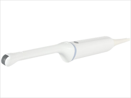
Featured probes for Voluson P8
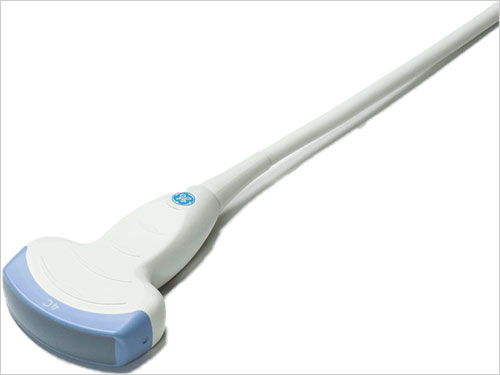
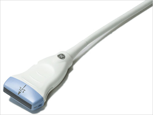
| 4C-RS curved probe | 12L-RS linear probe | IC9-RS endovaginal probe |
| Abdominal probe for excellent image quality for obstetric exams. | The 12L-RS linear probe features a high-performance broadband probe with a broad frequency range of 4-12 MHz and scanning range of 37 mm. This probe technology guarantees particularly fine, artifact-free imaging and the highest colour Doppler sensitivity. | Endovaginal probe for outstanding 2D gynecology and fetal imaging |
 |
 |
 |
Voluson P8: full probe list
|
|